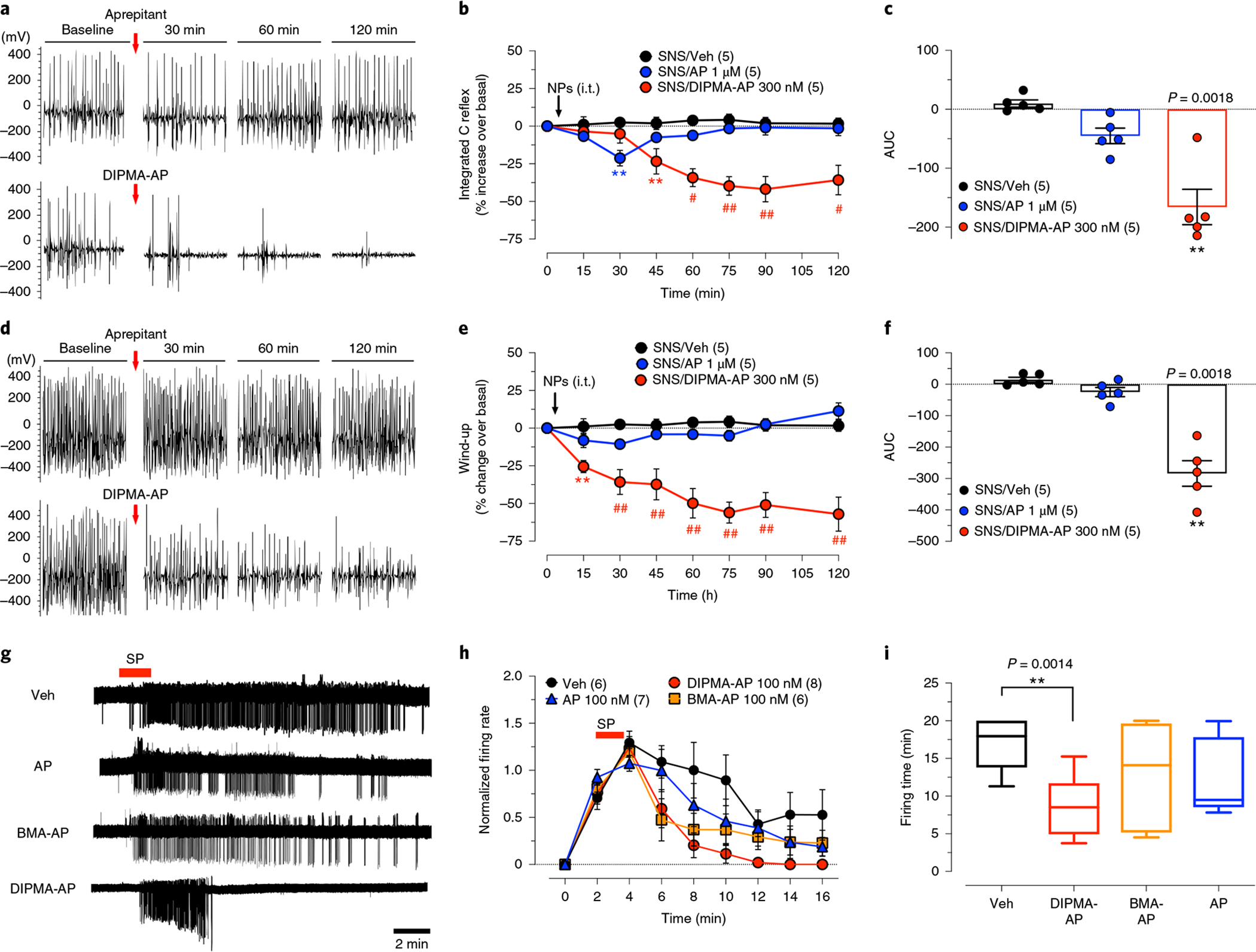
Fig. 5 |. Sensitization and activation of nociceptive transmission.

a–f, C-fibre reflex and wind-up in SNS rats. C-fibre reflexes (a–c) and wind-up (d–f) were measured at 10 days after SNS. AP, DIPMA-AP NP or Veh was administered by i.t. injection (10 μl). a,d, Representative recordings comparing AP and DIPMA-AP. b,e, Time course of effects. Data are presented as mean ± s.e.m., n = 5 rats per group (in parentheses). **P < 0.005, #P < 0.001, ##P < 0.0001 compared to vehicle. Two-way ANOVA, Dunn’s post-hoc test. c,f, Integrated responses (AUC, n = 5 rats). **P < 0.005, vehicle compared to DIPMA-AP, one-way ANOVA, Dunn’s post-hoc test. g–i, Cell-attached patch-clamp recordings of SP-induced excitation of lamina I neurons in slices of rat spinal cord. Tissues were preincubated with AP, NP or Veh, and then superfused with SP (1 μM, 2 min). Action potential firing was measured: representative traces (g); normalized firing rate (h); firing time (i). Data are presented as mean ± s.e.m., n = 6 for rats for Veh, n = 7 rats for AP, n = 8 rats for DIPMA-AP and n = 6 rats for BMA-AP. **P = 0.005, vehicle compared to DIPMA-AP. Unpaired t-test (two-sided).
