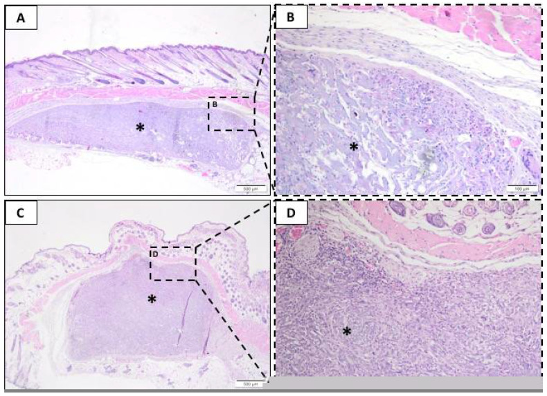
Figure 3.
Representative photomicrographs of subcutaneous injury after 21 days. Histological section stained with hematoxylin/eosin from the incision region in the BG groups (A,B) and LT groups (C,D). The area occupied by the membrane is indicated by an asterisk (*). (A,C) 40× magnification, scale bar: 500 µm; (B,D) 200× magnification, scale bar: 100 µm.

