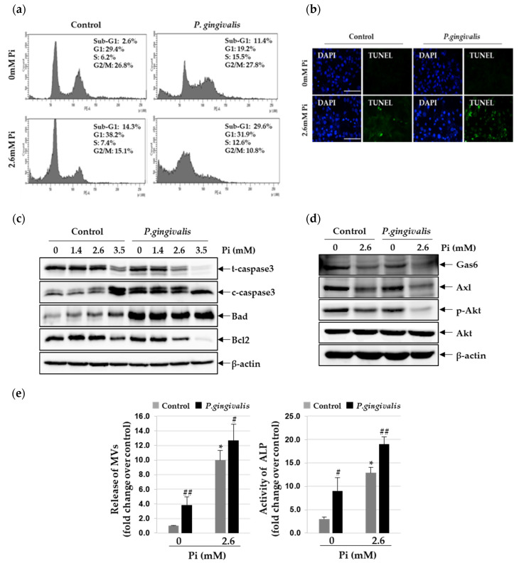
Figure 3.
Effect of P. gingivalis on Pi-induced apoptosis and matrix vesicle release in VSMCs. P. gingivalis-infected A7r5 cells were cultured in calcification medium (2.6 mM Pi) for three days. (a) Induction of apoptosis was determined by flow cytometry analysis with PI staining. (b) Apoptosis-associated DNA fragmentation was detected by fluorescent TUNEL (green), and cell nuclei were stained by DAPI (blue). (original magnification, ×400). Scale bar: 50 μm. (c) Total/cleaved-caspase 3, Bcl2, Bad, and β-actin protein levels were examined by Western blotting using their corresponding antibodies. (d) Gas6, Axl, p-Akt, Akt, and β-actin protein levels were examined by Western blotting using their corresponding antibodies. (e) Matrix vesicles were isolated as described in the Materials and methods section. Alkaline phosphatase activity was measured and normalized to the total protein content of matrix vesicles. * p < 0.01 vs. 0 mM Pi, # p < 0.05; ## p < 0.01 vs. control, one-way ANOVA followed by a Student’s t-test. Data shown are the mean ± SD, obtained for at least three independent experiments.

