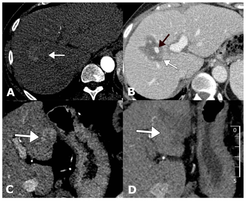
Figure 3.
Examples of tumors located centrally and peripherally before and after treatment with ECT. Arterial-phase contrast-enhanced computed tomography (CECT) demonstrates centrally located HCC with a diameter of 31 mm located in the vicinity of a major portal vein branch (A). Follow-up CECT 1 month after ECT demonstrates a complete response with no tumor enhancement and a patent portal vein branch (B). Arterial-phase CECT demonstrates peripherally located HCC in liver segment III (C). Follow-up CECT 1 month after ECT demonstrates a complete response with no tumor enhancement (D).

