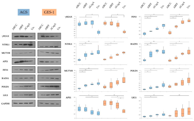
Figure 7.
Expression of DNA damage repair components in H. pylori-infected AGS (blue) and GES-1 (orange) cells. Results suggest CagA-independent increase of Ser139 phosphorylated histone H2AX (γH2AX) and decrease of Nth Like DNA Glycosylase 1 (NTHL1), MutY DNA Glycosylase (MUTYH), Flap Structure-Specific Endonuclease 1 (FEN1), RAD51 Recombinase, DNA Polymerase Delta Catalytic Subunit (POLD1), and DNA Ligase 1 (LIG1) protein levels, related to the expression and phosphorylation of CagA. Apurinic/Apyrimidinic Endodeoxyribonuclease 1 (APE1) protein levels were increased during the infection in a CagA-related manner. Quantification of protein levels was conducted by densitometry in at least three experimental replicates per condition. Statistical analysis was performed using Mann–Whitney U test (levels of significance: + p = 0.1–0.05, * p = 0.05–0.01, ** p < 0.01); Un.: uninfected control.

