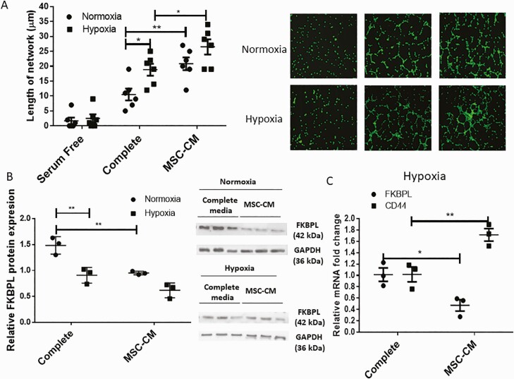
Figure 4.
Angiogenic properties of HUVECs are improved as a result of hypoxia and mesenchymal stem cell treatment, in association with reduced FKBPL and increased CD44 expression. (A) HUVEC differentiation was evaluated using a tubule formation assay; cells were stained with calcein and trypsinized before plating 50 000 cells in Matrigel and exposing them to hypoxia (1% O2) or normoxia (21% O2) for 6 h in complete or MSC-CM medium (n = 6). Cells were imaged using a Leica DMi8 fluorescent microscope and the tubules quantified using ImageJ software. Representative pictures are shown in the inset. (B) Protein lysates were collected and Western blotting performed to determine FKBPL (42 kDa) and glyceraldehyde 3-phosphate dehydrogenase (37kDa) protein expressions (n = 3). FKBPL protein expression was quantified using ImageJ and adjusted to glyceraldehyde 3-phosphate dehydrogenase expression. A representative picture of the Western blot is shown in the inset. (C) Real-time qPCR analyses of FKBPL and CD44 mRNA expression relative to the 18S housekeeping gene were carried out using HUVECs exposed to complete or MSC-CM in hypoxia for 24 h (n = 3). Results are expressed as mean ± SEM; statistical analyses were performed using 2-way ANOVA with Sidak’s or Tukey’s multiple comparisons test. *P < 0.05, **P < 0.01. Abbreviation: MSC-CM, mesenchymal stem cell–conditioned medium.

