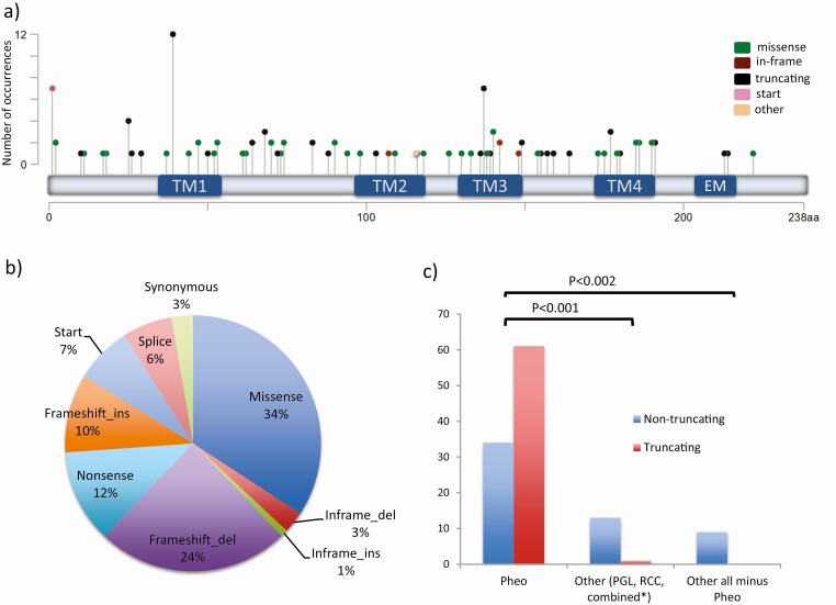
Figure 1.
Distribution of transmembrane protein 127 gene (TMEM127) variants. A, Diagram of the 111 germline TMEM127 variants along the amino acid sequence, displayed as lollipop symbols designed using the Mutation Mapper tool of the cBioPortal website (45, 46) and color coded based on the mutation class. The Y axis represents the number of occurrences of each variant; TM, transmembrane domains; EM, endocytic motif (19). B, Distribution of the 111 TMEM127 variants based on the mutation class (% shown). C, Distribution of TMEM127 variants (truncating vs nontruncating) based on clinical presentation into 3 groups: pheochromocytoma (Pheo) only; paraganglioma (PGL), renal cell carcinoma (RCC), either alone or combined with Pheo; PGL, RCC without Pheo; truncating variants include: nonsense, frameshift indels, splice site, and start site variants; nontruncating include missense and in-frame insertions or deletions.

