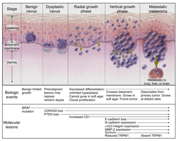
Figure 4.
Melanoma progression diagram (reproduced with permission from the New England Journal of Medicine, Arlo J. Miller & Martin C. Mihm, “Melanoma,” 355: 51–65, Copyright © 2020 Massachusetts Medical Society (Waltham, MA, USA). Reprinted with permission from Massachusetts Medical Society, [32]). Benign nevi have been known to express BRAF mutations that allows for benign and limited growth. This mutation results in the activation of mitogen-activated protein pathway. In the evolution to a dysplastic nevus, cytologic atypia becomes more prevalent through the loss of cyclin dependent kinase inhibitor 2A (CDKN2A) and phosphatase and tensin homologue (PTEN), resulting in what is often referred to as a premalignant lesion. The radial growth phase shows clonal proliferation and decreased differentiation accompanied by decreased expression of MITF (micropthalmia-associated transcription factor). The formation of a tumor characterizes the vertical growth phase with the crossing of the basement membrane. An absence of TRPM1 (melanocyte-specific gene melastitin 1) correlates with metastatic capability, but the function of the gene is unknown. There are several other genes involved in the melanoma including loss of E-cadherin, increased expression of N-cadherin, αVβ3 integrin expression, and survivin.

