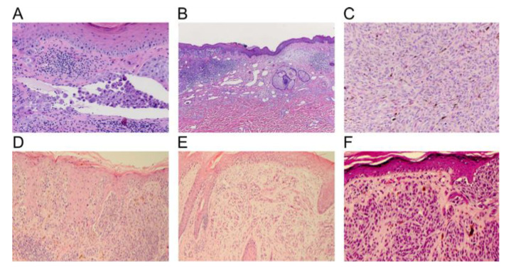
Figure 7.
Histopathological melanoma features. (A, B, and C reproduced from “Progress in melanoma histopathology and diagnosis” by Piris et al. with permission from the publisher Elsevier, Copyright © 2020 Elsevier Inc. (Amsterdam, The Netherlands) All rights reserved [45]) (A) Vascular invasion of malignant cells, (B) Lower-power view demonstrating partial regression of malignant melanoma, (C) dermal mitotic figures with dark blue pycnotic nuclei [45], (D–F) reprinted with permission from “The classification of cutaneous melanoma” by LM Duncan with permission from the publisher Elsevier, Copyright © 2020 Elsevier Inc. All rights reserved [46]. (D) Intraepidermal tumor cells within a superficial spreading melanoma with nests of cells and individual cell scatter present. (E) Intraepidermal tumor cells in a lentigo maligna melanoma that are located at the base of the epidermis with extension down the follicle of hair, and with the presence of epidermal atrophy. (F) Nodular melanoma with minimal tumor within the epidermis with nested proliferations of melanoma cells [46].

