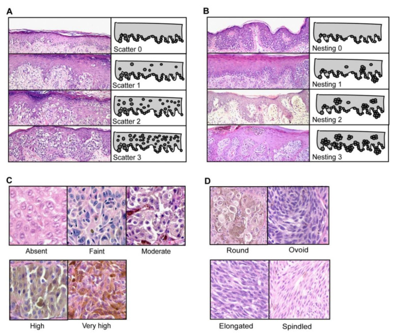
Figure 8.
Grading of cellular morphological features (Reproduced from “Improving melanoma classification by integrating genetic and morphologic features” by Viros et al. under Creative Commons Attribution License from PLOS Med, © Viros 2008 et al. [47]). (A) Shows grading of scatter of intraepidermal melanocytes from 0 to 3, increasing in scatter. Grade 0 shows all melanocytes along the dermal–epidermal junction; grade 1 shows >75% of melanocytes along the dermo-epidermal junction, with some present higher in the epidermis; Grade 2 showed equal amounts of intraepidermal melanocytes at the junction and higher in the epidermis; Grade 3 is noted when >50% of intraepidermal melanocytes are in the upper epidermis, (B) shows the grading of nesting of intraepidermal melanocytes from 0 to 3. The amount of nesting is quantified here as: Grade 0: Intraepidermal melanocytes present as single cells with rare nests; Grade 1: Intraepidermal melanocytes arranged as single cells with <25% of cells in nests; Grade 2 Intraepidermal melanocytes in nests in 25%–50%; Grade 3: >50% of intraepidermal melanocyte population are arranged in nests, (C) Cytoplasmic pigmentation of neoplastic melanocytes, Scaled 0–4. Score of 0 meant no pigmentation is present; Score 1: Faint pigmentation barely visible at low power; Score 2: Moderate pigmentation visualized at low power; Score 3: High pigmentation easily visible at low power, pigmentation of cytoplasm is similar to the nucleus pigmentation intensity; Score 4: Very highly pigmented cytoplasm, often obscuring nuclei [47]. (D) Cell shapes were also graded 0–3, Grade 0: Round cell with equal diameter and length; Grade 1 was ovoid with a diameter 1/3 longer than the short diameter; Grade 2 is elongated with a long diameter 1/3–2 times longer than the short diameter; Grade 3 is spindled with a long diameter two times the shorter diameter [47].

