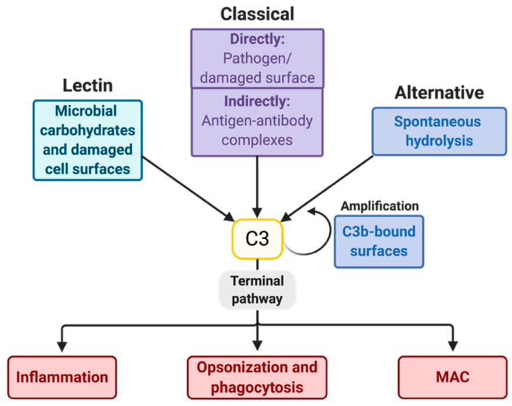
Figure 1.
Pathways of the complement system. Many surfaces of pathogens, immune complexes, or modified host surfaces (e.g., necrotic and apoptotic cells [10,11]) activate the complement system. In the lectin pathway (LP), microbial carbohydrates or modified self surfaces [12] are recognized by lectins, either by one of the two collectins, mannose-binding lectin (MBL) or collectin-LK, or by one of three ficolins [13]. C1q is the recognition component of the classical pathway (CP) and recognizes either deposited immunoglobulins (Igs) or C-reactive protein [14], or binds the pathogen or apoptotic cell directly [10]. Complement component 3 (C3) may also be activated through spontaneous hydrolysis of its thioester in the alternative pathway (AP). The AP can also amplify C3-responses via the formation of a C3-convertase. The cleavage of C3 initiates the terminal pathway (TP), which introduces inflammation, opsonization, and subsequent phagocytosis as well as the formation of the membrane-attack complex (MAC). The MAC stimulates certain signaling pathways [15,16,17,18] or lyses cells to disturb their integrity. AP, alternative pathway. C3, complement component 3. CP, classical pathway. Ig, immunoglobulin. LP, lectin pathway. MAC, membrane-attack complex. TP, terminal pathway.

