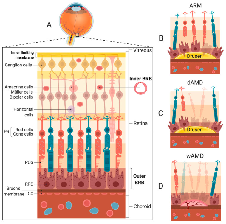
Figure 2.
The cellular organization of the retina, age-related maculopathy (ARM), and age-related macular degeneration (AMD) stages. (A) The different cell layers of the retina are depicted spanning from the vitreous body to the choroid. The outer layer of the retina comprises photoreceptor (PR) cells and the retina pigment epithelium (RPE). Bruch’s membrane stretches from the plasma membrane of the RPE to the choriocapillaris (CC) in the choroid, which is the vascularized layer of the eye comprising vessels and connective tissue. The inner limiting membrane is the boundary between the retina and the vitreous formed by Müller cell footplates. (B) Progression of the healthy retina to age-related maculopathy (ARM) is ascertained by the presence of drusen and/or abnormal pigmentation. (C) Geographic atrophy (GA) is distinguished by one or more sharply delineated areas of at least 175 µm in diameter with abnormal pigmentation or local areas of complete RPE atrophy and more visible choroidal vessels than the surrounding areas. (D) wet AMD (wAMD) is ascertained when RPE detachment and/or choroidal neovascularization (CNV) is present. Acute vision loss can be experienced due to the accumulation of subretinal and/or intraretinal fluid with a loss of structural and functional retinal integrity. ARM, age-related maculopathy. BRB, blood-retina barrier. CC, choriocapillaris. CNV, choroidal neovascularization. dAMD, dry AMD. GA, geographic atrophy. POS, photoreceptor outer segment. PR, photoreceptor. RPE, retinal pigment epithelium. wAMD, wet AMD.

