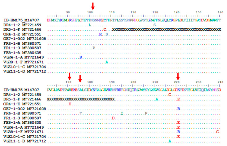
Figure 5.
Alignment of the deduced amino acid sequences of the VP1 region of the HM175 strain and hepatitis A virus (HAV) strains carrying mutations at immunodominant and neutralisation epitopes. Only amino acid position 81 to 240 is shown. Conserved sites, substitutions and deletions are represented by dots, single-letter abbreviation and the letter “X”, respectively. The red arrows point to the positions of the immunodominant (102, 171 and 176) and neutralisation (221) epitopes. The complete protein alignment from position 1 to 300 is provided as Supplementary files 3 (graphic view) and 4 (Fasta file).

