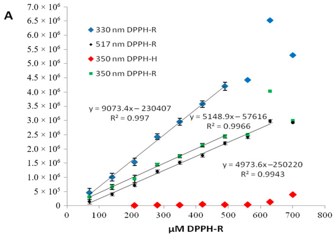
Figure 3.
Dependence of the peak area of the DPPH radical on its concentration measured at different wavelength. Chromatographic conditions: column—a Zorbax Eclipse XDB-C18 (4.6 × 150 mm, 5 μm), mobile phase—methanol/water (80:30, v/v), flow rate—1 mL/min. The presence of the trace content of DPPH-H (2,2-diphenyl-1-picrylhydrazine) at each measured sample is illustrated in red.

