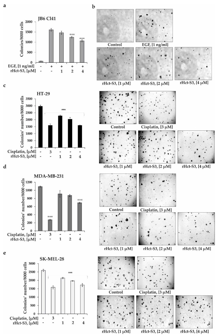
Figure 3.
Effect of rHct-S3 on EGF-induced neoplastic cells transformation of JB6 Cl41 cells and colony formation of human colorectal carcinoma HT-29, breast cancer MDA-MB-231, and melanoma SK-MEL-28 cell lines. (a,b) JB6 Cl41 cells (2.4 × 10⁴ /mL) treated with/without EGF (1 ng/mL) or investigated compound (1, 2, and 4 µM) in 1 mL of 0.3% Basal medium Eagle (BME) agar containing 10% FBS and overlaid with 3.5 mL of 0.5% BME’s agar containing 10% FBS. The culture was maintained at 37 °C in a 5% CO2 atmosphere for 2 weeks. (c) HT-29, (d) MDA-MB-231, (e) SK-MEL-28 cells (2.4 × 10⁴ /mL) treated with/without investigated compound (1, 2, and 4 µM) or cisplatin at 3 µM (positive control) and subjected into a soft agar. The culture was maintained at 37 °C in a 5% CO2 atmosphere for 2 weeks. The colonies were counted under a microscope with the aid of the ImageJ software program (n = 6 for control and each compound, n—quantity of photos). The magnification of representative photos of colonies is ×10. The asterisks (*** p < 0.001) indicate a significant decrease in colony formation in cells treated with compound compared with the non-treated cells (control).

