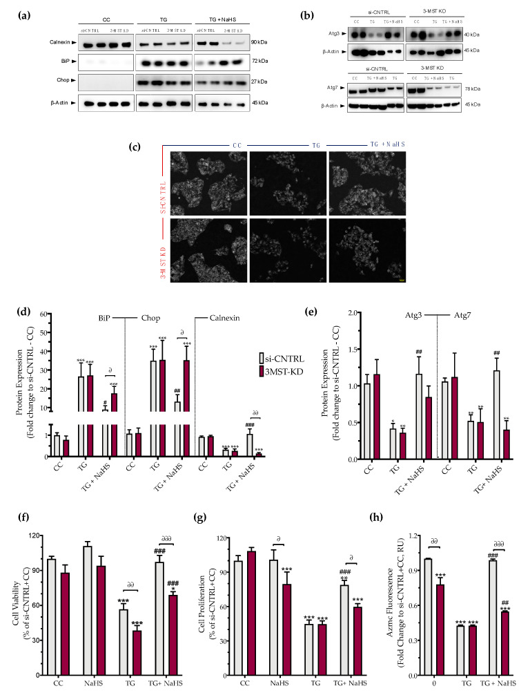
Figure 9.
3-MST silencing attenuates the restorative effects of the H2S donor NaHS on the ER stress response and autophagy. HepG2 cells previously subjected to 3-MST silencing (knockdown (KD)) or sham-silencing (si-CNTRL) were serum-starved for 8 h and treated with the vehicle or 100 nM of thapsigargin (TG), in the presence or absence of 100 µM NaHS for 16 h. Whole-cell lysate was collected and processed for protein expression of the UPR sensor–BiP, the UPR apoptotic mediator–Chop, the ER chaperone–calnexin (a,d), and of the autophagy-related proteins Atg3 and Atg7 (b,e). Representative images of the cells previously labeled with the Autophagosome Detection probe were captured under the Olympus CKX53 inverted fluorescent microscope with a DAPI channel. Autophagy is indicated by bright dot staining of autophagic vacuoles (c). Micro-cultures were assayed for XTT conversion (f) BrdU incorporation (g), and AzMC fluorochrome intensity (h) to assess cell viability, cell proliferation, and intracellular H2S levels, respectively. Each bar represents mean ± SEM from three and four independent experiments for Western blotting and cell-physiology studies, respectively. Data are expressed as a percentage of the transfection negative control, control (vehicle-treated) conditions (si-CNTRL+CC). * p ≤ 0.05, ** p ≤ 0.01 and *** p ≤ 0.001 compared to corresponding CC; # p ≤ 0.05, ## p ≤ 0.01 and ### p ≤ 0.001 compared to the corresponding TG-treated cells; ∂ p ≤ 0.05, ∂∂ p ≤ 0.01 and ∂∂∂ p ≤ 0.001 compared to the corresponding 3-MST KD treatment group.

