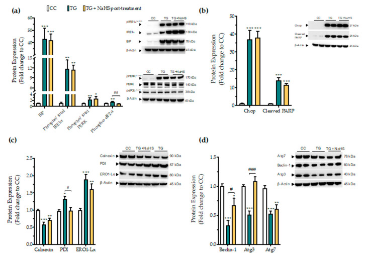
Figure 12.
Delayed exogenous supplementation with H2S does not ameliorate UPR activation though it partially rescues autophagy impairments. HepG2 cells were serum-starved for 8 h and treated with the vehicle or 100 nM thapsigargin (TG) for 16 h. NaHS treatment initiated 4 h post thapsigargin. Next, cells were harvested and the extracted protein was processed for Western blotting analysis of the expression levels of BiP, activating phosphorylation of IRE1α at the serine residue 724 (Ser724), the activating phosphorylation of PERK at the threonine residue 980 (Thr980), the inhibitory phosphorylation of eIF2α at Ser51 (a). We additionally quantified the expression levels of the UPR apoptotic mediator Chop and of the cleaved PARP (b). Moreover, the expression of the chaperones–calnexin, PDI and ERO1-Lα (c), and of the autophagy-related proteins (Atg)-beclin-1, Atg3 and Atg7 (d) were assessed. Each bar represents mean ± SEM from three independent experiments. Data are expressed as a fold change of the control (vehicle-treated) conditions (CC). * p ≤ 0.05, ** p ≤ 0.01; *** p ≤ 0.001 compared to CC; # p ≤ 0.05, ## p ≤ 0.01, ### p ≤ 0.001 compared to TG-treated cells. For the abbreviations used, please refer to the main text.

