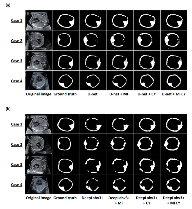
Figure 2.
Representative examples of the thoracic wall segmentation by the existing models and our three methods on test datasets. Each row shows a particular case. The columns correspond to the original images, ground truths, and predictions by U-net, DeepLabv3+, MF, CY, and MFCY. The white areas represent the labels of the thoracic wall; (a) corresponds to U-net and (b) corresponds to DeepLabv3+.

