Figure 1.
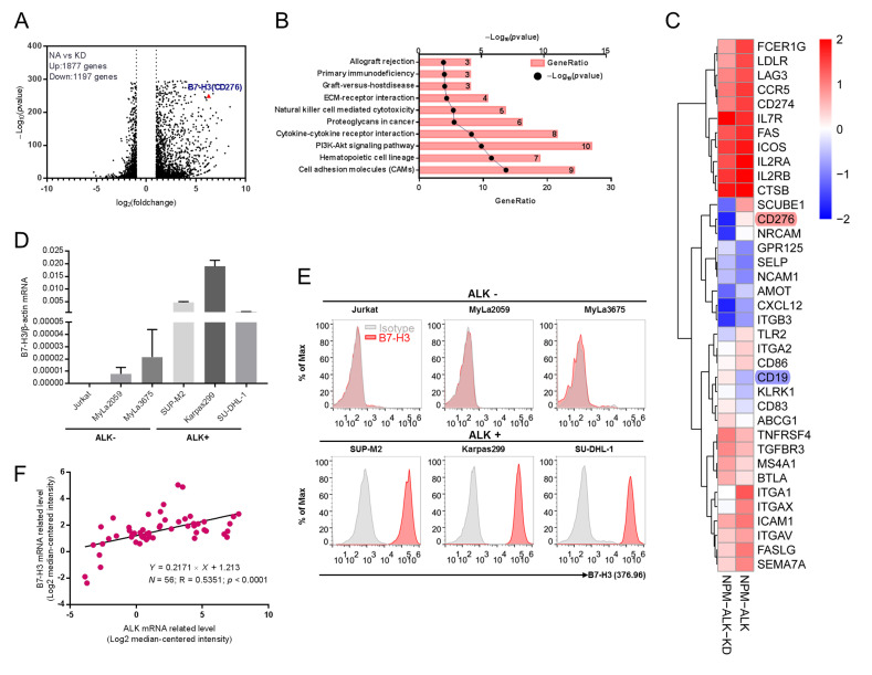
Overexpression of B7-H3 is detected in ALK+ ALCL. (A) Purified CD4+ T cells were stimulated with a T cell expander (bead-immobilized CD3 and CD28 antibodies) and either transduced with wild-type NPM-ALK (NA) or enzymatically inactive NPM-ALK mutant (KD). RNA was isolated on day 10 after transduction and deep sequencing was performed. Volcano plots depict differently expressed genes (DEGs) in NA and KD transduced CD4+ T cells. B7-H3 (CD276) is the red triangle. (B) Pathway enrichment analysis of different expressed membrane proteins (DEMGs) in NA vs. KD transduced CD4+ T cells. The top 10 pathways were presented. (C) The top 37 altered DEMGs between the NA and KD transduced CD4+ T cells group were presented in the heatmap. (D) B7-H3 mRNA level was analyzed by quantitative reverse-transcriptase PCR (qPCR) in both ALK-negative and ALK-positive cell lines. (E) B7-H3 expression on cell membrane was evaluated via flow cytometry analysis. Isotype control (grey graphs, isotype control rabbit IgG as primary antibody and anti-rabbit Fc as secondary antibody) and B7-H3 (red graphs, anti-B7-H3 antibody as primary antibody and anti Fc as secondary antibody). (F) B7-H3 expression positively correlates with ALK expression found on Oncomine database (n = 56, R = 0.5351).

