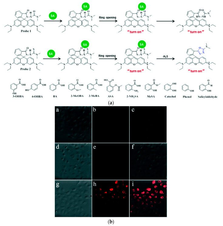
Figure 18.
Detection of salicylic acid: (a) Proposed mechanism for detecting SA using probe 1 (top) and 2 (middle) and structures of tested SA analogues (bottom); (b) bright-field (left) and fluorescence (middle) and merged (right) images of NRK-52E cells; control (top) probe 2 (middle) and probe 2 + SA (bottom). Adapted with permission from [244].

