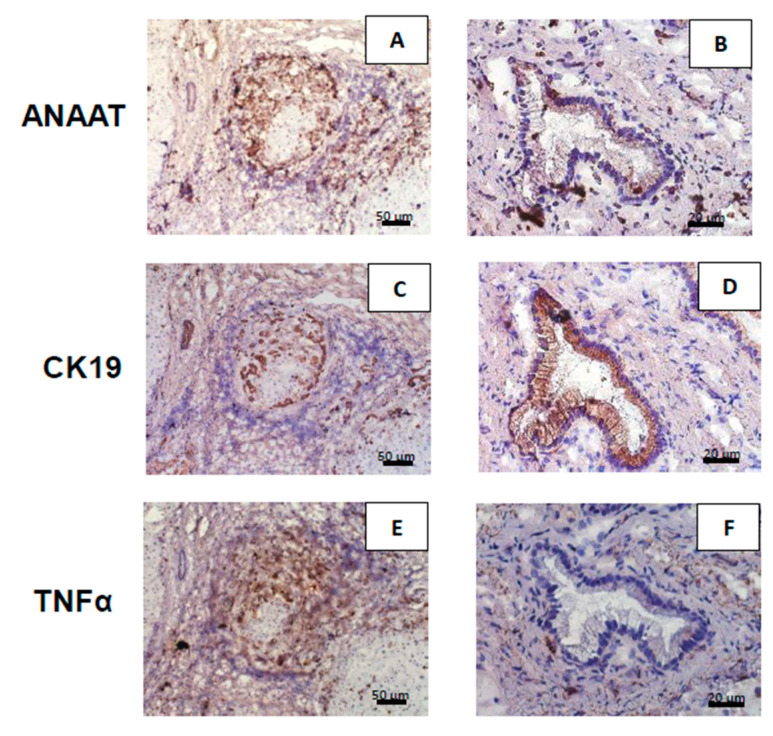
Figure 2.
Hepatic expressions of aralkylamine N-acetyltransferase (AANAT), CK19, and TNFα protein in primary biliary cholangitis (PBC) patients. Positive immunohistochemical staining (dark brown) of AANAT, CK19, and TNFα in cirrhotic liver tissues were observed on the edges of regenerative nodules (A,C,E), and in bile ducts (B,D,F), respectively. (A,B) AANAT was ubiquitous in the liver tissue of PBC patients. (C,D) CK19 was localized in cholangiocytes in areas of ductular reactions. Bile ducts are marked by asterisks. (E,F) TNFα was localized primarily on the perimeter of nodules, but was absent in the cholangiocytes of the large bile duct.

