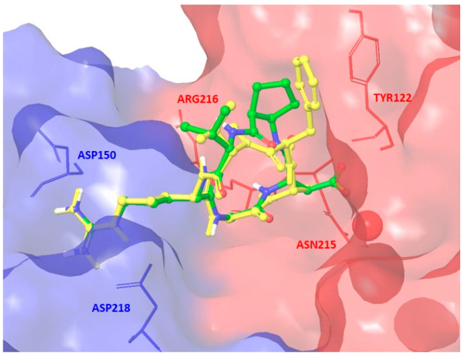
Figure 5.
Docking best pose of compound 8 (Type I, green C atoms, Gscore = −9.26 kcal/mol) in the crystal structure of the extracellular domain of αvβ3 integrin (α unit blue, β unit red) overlaid on the bound conformation of Cilengitide (yellow). Only selected integrin residues involved in interactions with the ligand are shown. The metal ion at MIDAS is shown as a red CPK sphere (space-filling model by Corey, Pauling, Koltun). Nonpolar hydrogens are hidden for clarity.

