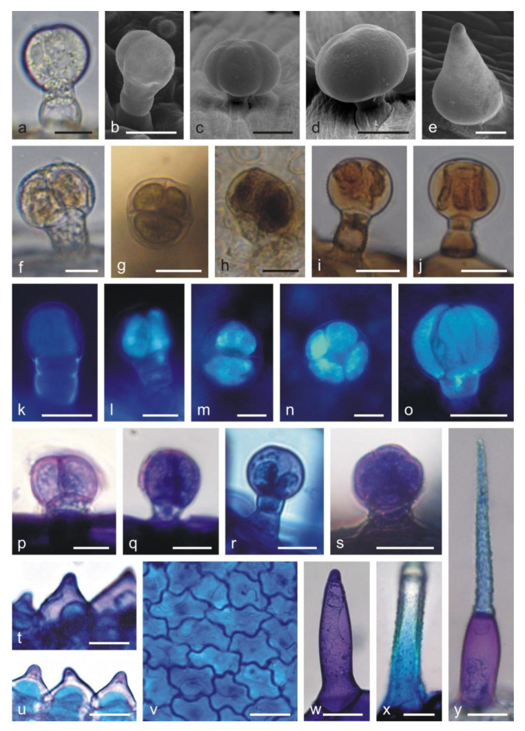
Figure 2.
Types of trichomes and papillae on the L. album subsp. album corolla and the results of histochemical assays. (a,f) Control trichomes without staining (LM). (b–e) SEM images of trichomes. (g–j,p–y) Histochemical reactions (LM). (k–o) fluorescence microscopy (FM) images of trichomes. (a) Capitate trichome with a 1-celled head. (b) Capitate trichome with a 2-celled head. (c) Capitate trichome with a 4-celled head. (d) Peltate trichome. (e) Conical trichome. (f) Capitate trichome with a bicellular head and yellow content in the head cells. (g,h) Phenolic compounds stained black in the heads of trichomes after the application of ferric trichloride. (g) Capitate trichome with a 3-celled head. (h) Peltate trichome. (i,j) Brown color of tannins in trichomes stained with potassium dichromate. (k–o) Blue autofluorescence in different capitate and peltate (o) trichomes indicating the presence of phenolic acids. (p–r) Tannins in capitate trichomes stained blue after Toluidine blue O treatment. (s) Peltate trichome stained blue after Toluidine blue O treatment. (t–u) Phenolic compounds in papillae stained blue after Toluidine blue O treatment. (v) Phenolic compounds visible in abaxial epidermis cells of the upper lip after Toluidine blue O treatment. (w) Non-glandular conical trichome from the corolla tube stained purple (pectins) after Toluidine blue O treatment. (x,y) Non-glandular trichomes containing phenolic compounds (blue) visible after Toluidine blue O treatment. Scale bars: 30 µm (c,d,h,i,q,r,w,y), 20 µm (a,b,f,g,j,k,o,p,s,t,u,v), 10 µm (e,l,m,n,x).

