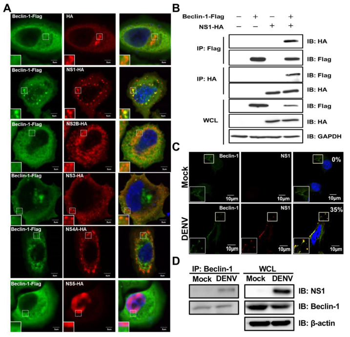
Figure 4.
The colocalization and interaction of DENV NS1 with Beclin-1 are found in NS1-expressing and DENV-infected cells. (A) Hela cells were co-transfected with NS plasmids (including HA, NS1-HA, NS2B-HA, NS3-HA, NS4A-HA or NS5-HA) and Belcin-1-Flag plasmids for 48 h. At 48 h post-transfection, cells were fixed, permeabilized, and stained with anti-HA Abs (green), anti-Flag Abs (red) and DAPI (blue). Cells were mounted and observed by confocal microscopy. The square insets show higher magnification. Scale bar: 5 μm. (B) Hela cells were co-transfected with NS1-HA and Belcin-1-Flag plasmids for 48 h. Cells were lysed and the cell lysates were immunoprecipitated by anti-Flag Abs followed by immunoblotting using anti-HA and anti-Flag Abs. The whole cell lysate (WCL) was used as the protein control. (C) A549 cells were infected with DENV at MOI = 2 for 24 h. At 24 h post-infection, cells were fixed, permeabilized, and stained with anti-NS1 Abs (green), anti-Beclin-1 Abs (red) and DAPI (blue). Cells were mounted and observed by confocal microscopy. The square insets show higher magnification. Percentages of colocalized dots of NS1-Beclin-1 complex were indicated in the upper right corner. Scale bar: 10 μm. (D) Cells were lysed and the cell lysates were immunoprecipitated by anti-Beclin-1 Abs followed by immunoblotting using anti-NS1 and anti-Beclin-1 Abs. The whole cell lysate (WCL) was used as the protein control.

