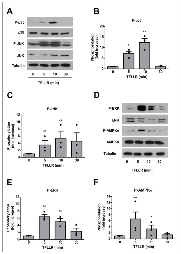
Figure 4.
PAR1 activation in BBB endothelial cells leads to MAPK and AMPK phosphorylation. GPNT were stimulated with 100 µM TFLLR for the indicated length of time, and p38 (A,B), JNK (A,C), ERK (D,E) and AMPKα (D,F) phosphorylation was assessed by western blot. Representative results and quantification of protein phosphorylation, normalised to total protein levels, from three independent experiments are shown as means ± SEM. Each data point represents one independent, biological replicate. Significant differences from controls were determined by ANOVA and Dunnett’s post-hoc analysis with * p < 0.05, ** p < 0.01.

