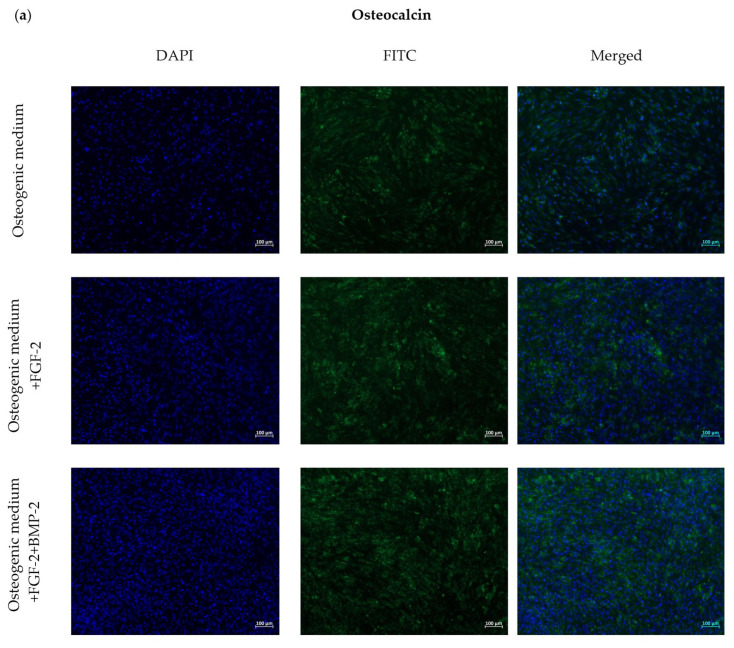
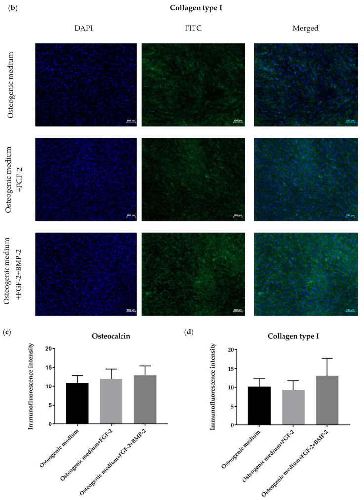
Figure 5.
Representative images for the immunofluorescence staining of osteocalcin (Ocl) (a) and collagen type I (ColI) (b), expressed by BM-MSCs cultured in an osteogenic differentiation medium with or without BMP-2 and/or FGF-2 for 21 days. The Ocl and ColI were stained with FITC in green and nuclei in blue with DAPI. Immunofluorescence staining for osteocalcin (c) and collagen type I (d) was quantified using the ImageJ software. FGF-2 and BMP-2 promoted the expression of osteogenic-related proteins, which resulted in a moderately more intense fluorescence in the treated cells compared to the control cells cultured in the osteogenic differentiation medium alone.


