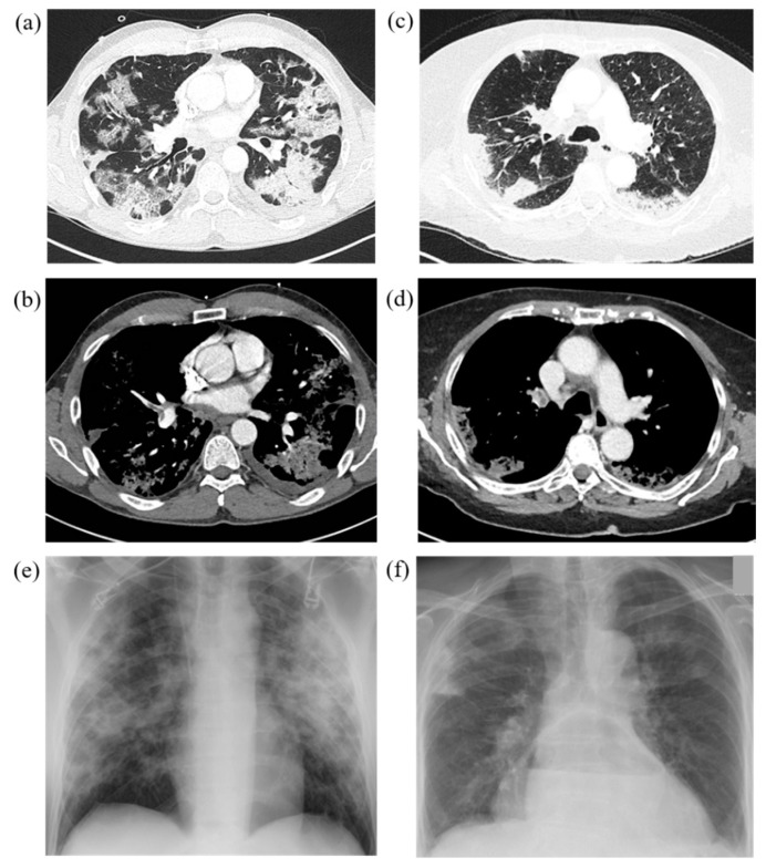
Figure 3.
Representative cases with typical COVID-19 findings on chest CT and chest radiograph in both study groups. IV = Invasive ventilation group; non-IV = No/non-invasive ventilation group. (a)/(b) Chest CT of a 49-year-old, PCR-confirmed male COVID-19 patient in the IV-group after onset of symptoms 15 days ago with immobilization due to reduced general condition and fever, dry cough and headache. CT on admission reveals GGOs, crazy paving pattern, consolidations, subpleural bands in the lower lobes, lymph node enlargements and right central pulmonary embolism. IV risk-score on admission was 7 points out of 10. (c)/(d) 81-year-old female COVID-19 patient in the non-IV-group with onset of symptoms one week ago presenting with fever, dry cough and reduced general condition, diarrhea and nausea. Central pulmonary embolism in the both pulmonary arteries is noted. GGOs as well as consolidations were observed, partially rated as pulmonary infarction. IV-risk score on admission was 1 point out of 10. Thoracic stomach as secondary finding. (e)/(f) Corresponding chest radiographs of both cases showed signs of atypical pneumonia as well as pulmonary infarction due to pulmonary embolism.

