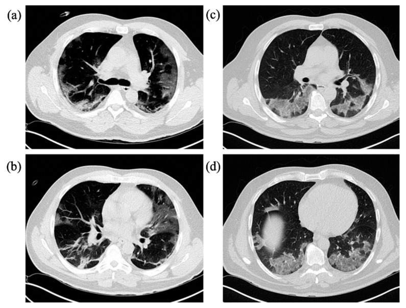
Figure 5.
Representative cases with typical COVID-19 imaging features on chest CT in a group comparison. IV = Invasive ventilation group; non-IV = No/non-invasive ventilation group. (a)/(b) 45-year-old male patient of the IV-group with known contact to a positively tested working colleague. Symptom onset was around 2 weeks earlier and worsened over the last week prior to admission with fever, sore throat and worsening general condition, progressive dyspnea in the preceding four days. IV-risk score of the patient on admission was 5 points with 5 lobes involved. GGOs as well as subpleural bands were the predominant finding in CT scan on admission. (c)/(d) 59-year-old male patient in the non-IV-group; no known exposure, beginning of symptoms about a week ago with fever, dry cough and reduced general condition. IV-risk score on admission was 2 with 5 lobes involved. GGOs was the predominant finding with some interseptal thickening within the regions, no consolidations.

