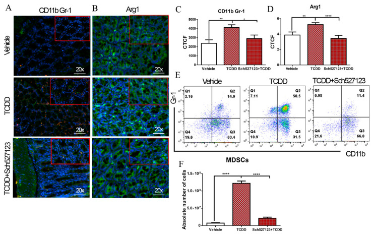
Figure 4.
Role of CXC chemokine receptor 2 (CXCR2) in TCDD-mediated induction of MDSCs. Colon samples from three groups (vehicle, TCDD, and TCDD with the CXCR2 antagonist Sch5271230; n = 6 per experimental group) were fixed with 4% paraformaldehyde in PBS overnight and then sectioned to 5 µm thickness on coated slides. Slides were incubated with antibody detection of Gr-1, CD11b, and Arg1. (A) CD11b (green) and Gr-1 (red) fluorescence staining in the colon to detect MDSCs (green + red overlap = yellow). 20x, scale bar = 50 µm. (B) Arg1 fluorescence staining (green) to quantify Arg1 expression in the colon of three groups. 20x, scale bar = 50 µm (C,D) Statistical analysis of CD11b, Gr-1, and Arg1 expression in colon sections of three groups as measured in corrected total cell fluorescence (CTCF) using ImageJ software. (E) Representative flow plots of MDSCs in peritoneal fluid in vehicle, TCDD, and TCDD with the CXCR2 antagonist Sch527123 groups. (F) Absolute numbers of MDSCs in peritoneal fluid in vehicle, TCDD, and TCDD with the CXCR2 antagonist Sch527123 groups. One-way analysis of variance (ANOVA) and Tukey’s multiple comparisons test were used to compare among the groups. * p < 0.05; ** p < 0.01; **** p <0.0001. Data are representative of at least two independent experiments.

