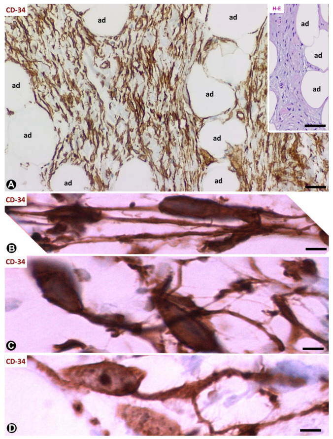
Figure 6.
Spindle cell lipoma. (A): CD34+ spindle cells (brown) are observed between adipose cells (ad). Insert shows an image with haematoxylin–eosin (HE) staining in which it is difficult to distinguish processes from spindle cells. (B–D): CD34+ spindle cells with bipolar processes (B), piriform aspect with a single (C) and branched (D) process. (A–D): CD34 immunochemistry; haematoxylin counterstain. Bar: (A): 80 µm; (insert of (A)): 100 µm; (B–D): 10 µm.

