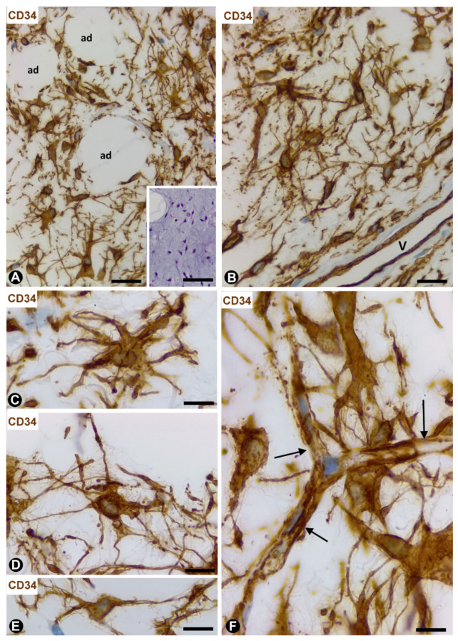
Figure 7.
Dendritic fibromyxolipoma. (A–F): CD34 immunochemistry with haematoxylin counterstain. CD34+ dendritic cells (brown) between adipocytes (ad) (A) and near a vessel (v, (B)). Observe the stellate aspect (C,D) and contacts with neighbouring cells (E) and small vessels ((F), arrows). Insert of A: image in a section stained with haematoxylin and eosin, bar: (A,B): 60 µm; (insert of (A)): 100 µm; (C–E): 20 µm; (F): 30 µm.

