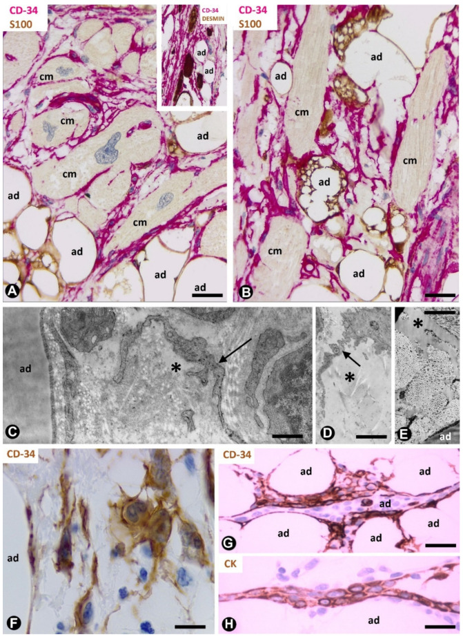
Figure 9.
TCs/CD34+SCs in the lipomatous hypertrophy of the interatrial septum, nevus cutaneous superficialis of Hoffman–Zurhelle and irradiated adipose tissue. (A,B): lipomatous hypertrophy of the interatrial septum. Double immunochemistry labelling for CD34 (red) and S100 protein (brown). Numerous TCs/CD34+SCs (red) are observed between cardiomyocytes (cm) and adipocytes (ad, brown). Note some multivacuolated cardiomyocytes. Insert of A: double immunochemistry labelling for CD34 (red) and desmin (brown). Desmin-positive cardiomyocytes (brown) are surrounded by TCs/CD34+SCs (red); adipocytes: ad. (C–E): nevus lipomatosus cutaneous superficialis of Hoffman–Zurhelle. Ultrastructural images of a TC ((D), arrow) or TC processes ((C), arrows) and free fat in the interstitium ((D,E), asterisks); adipocytes = ad. (F–H): irradiated fat in rectal and perirectal tissues (F) and in the thymic region (G,H). Immunochemistry labelling for CD34 (F,G) and cytokeratin (H). Increased numbers of TCs/CD34+SCs (brown) showing prominent processes. Observe some of these cells around inflammatory cells (F) and strips of thymic epithelium (G), which express cytokeratin ((H), brown). Bar: (A,B): 40 µm; (C–E): 4 µm; (F): 20 µm; (G,H): 40 µm.

