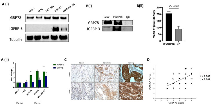
Figure 1.
GRP78 and IGFBP-3 co-operate (A,i) The levels of GRP78 and IGFBP-3 were examined using Western blotting of the whole cell lysates from the nonmalignant MCF-10A cells and various breast cancer cell lines. (A,ii) Quantification of Western blot results shown in (A,i). (B,i) Hs578T cell extract was subjected to IP with either control IgG or GRP78 antibodies and the samples immunoblotted with GRP78 and IGFBP-3 antibodies. Input represents whole cell lysate as a percentage of protein amount (12.5%) used for the IP reaction. (B,ii) Quantification of IP results presented in (B,i). (C) Representative images of GRP78 and IGFBP-3 IHC staining (1 = weak; 2 = moderate; 3 = strong) in paraffin-embedded breast cancer tissue sections (n = 69). IHC scores of GRP78 and IGFBP-3 abundance are shown in (D) Nonparameter Spearman correlation co-efficiency and the p value are also shown. The whole blots (uncropped blots) are shown in the Supplemental Materials.

