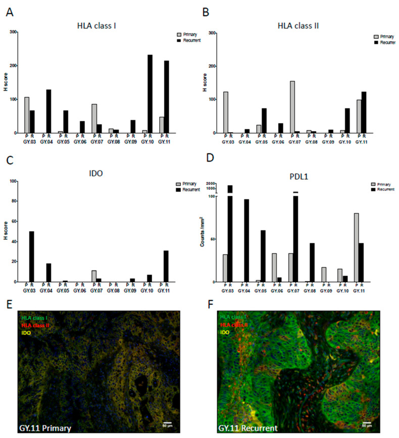
Figure 2.
The expression of immune-modulating molecules on tumor cells in the TME of the primary (P) and recurrent (R) tumor samples were analyzed using multispectral immunohistochemistry (IHC) analysis or immunofluorescent IHC. (A–D) expression in primary tumor is presented in light grey and expression in recurrent tumor is presented in black. (A) Expression of HLA class I in the epithelial compartment of the TME. (B) HLA class II in the epithelial compartment of the TME. (C) Expression of IDO in the epithelial compartment of the TME. (D) Expression of PDL1 (CD163-PD1-PDL1+). (E,F) Example of HLA and indoleamine 2,3-dioxygenase (IDO) expression in primary (E) and recurrent (F) tumor tissue in patient GY.11. HLA class I in green, HLA class II in red, and IDO in yellow.

