Figure 7.
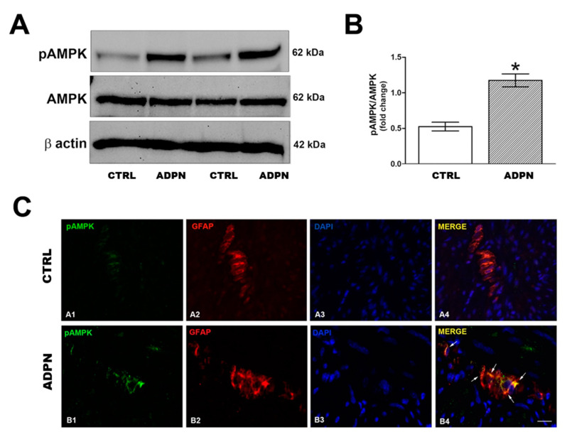
Involvement of AMPK signaling in ADPN action. (A) Effect of ADPN on AMPK signaling pathway in mouse gastric fundus assayed by Western blotting: representative bands from a typical experiment. (B) quantitative analysis. Columns are means ± SEM. Significance of differences (Student’s t-test for two independent samples): * p < 0.05 vs. controls (CTRL). (C) Representative photomicrographs of gastric tissue from control and ADPN-treated mice showing double immunofluorescence labeling. (A1–A4) co-localization of phosphoAMPK (pAMPK) and GFAP in control mice: (A1) pAMPK signal (green channel); (A2) GFAP signal (red channel); (A3) DAPI; (A4) merged images. (B1–B4) co-localization of pAMPK and GFAP in ADPN-treated mice: B1 pAMPK signal (green channel); (B2) GFAP signal (red channel); (B3) DAPI; (B4) merged images. Scale bar: 10 µm.

