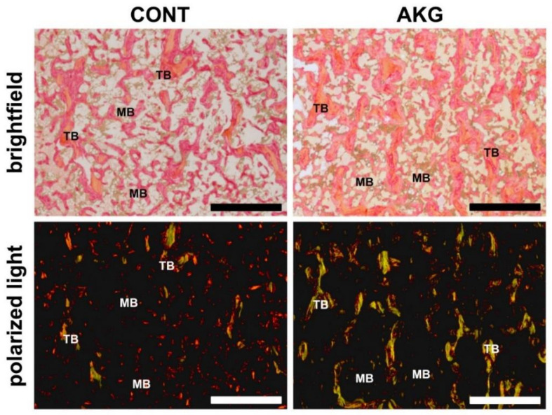
Figure 3.
The influence of AKG supplementation during the experimental period (31–60 weeks of age) on bone collagen in laying hens at the age of 60 weeks. Representative images of PSR-stained sections from the tibial trabecular (TB) and medullary (MB) bone of laying hens from the control (CONT) and AKG supplemented (AKG) groups, viewed with conventional brightfield light microscopy and polarized (polarizer and analyzer are “crossed”) light. The large mature collagen fibers are orange or red and the thick ones, including the reticular fibers, are green. All scale bars represent 100 μm.

