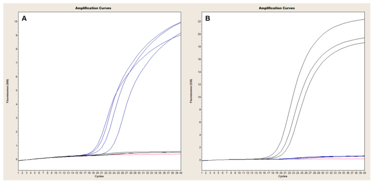
Figure 6.
Evaluation of capB-lef duplex PCR in LightCycler 2.0. A collection of B. anthracis and Bcbva strains were evaluated with the capB and lef TaqMan duplex PCR using 1 ng of genomic DNA (Table 4). Amplification of lef in capB-negative B. anthracis strains is illustrated in Panel (A) (blue signal), with little to no lef background signal detected in isolates lacking pXO1 (Panel (A), black signal). Conversely, capB signal was readily detected in strains harboring pXO2 (Panel (B), black signal), but absent from capB-negative isolates (Panel (B), blue signal).

