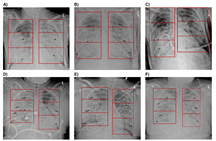
Figure 4.
Chest X-rays of patients with sustained improvement in PaO2/FiO2 and with decline in PaO2/FiO2 after 48 hours (h). Anterior-Posterior (AP) radiographs of the chest with lung zones demarcated by a red box. Images (A–C) are from a single patient with sustained improvement in PaO2/FiO2. Images (D–F) are from a single patient with a decline in PaO2/FiO2. (A) Image captured 48 h before proning demonstrates ground-glass opacities in the left and right upper lung zones (long arrows), with confluent consolidations in the right middle, right lower, left middle, and left lower zones (short arrows). Images (B) immediately after proning demonstrate improving infiltrates bilaterally with significant improvement in the left middle, left lower, and right lower lung zones, and (C) 48 h after proning demonstrate significant improvement in the infiltrates bilaterally with residual dense consolidation in the right middle and lower lung zones. (D) Image one day before proning demonstrates ground-glass opacities throughout all lung zones (long arrows), with diffusely scattered confluent consolidations bilaterally (short arrows). Images (E) immediately after proning demonstrate improving infiltrates, particularly in the right upper, left upper, and left middle lung zones, and (F) 48 h after proning demonstrate worsening of infiltrates bilaterally with increased confluent consolidations in the left and right upper lung zones.

