
FIGURE 5.
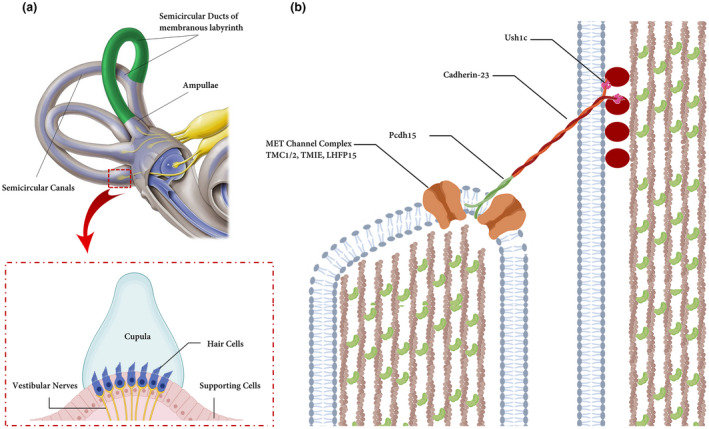
(a) Hair bundles and tip links. The diagram of a hair cell is depicted the hair bundle and the tip‐link filaments that connect the stereocilia in the direction of their mechanical sensitivity. Cupula is a structure in the vestibular system, providing a sense of spatial orientation. (b) Molecules form tip links and putative components of the mechanotransduction channels in hair cells. Cadherin‐23 interacts directly with protocadherin‐15 (Pcdh15) to form the upper and lower parts of tip links. Ush1c and Myosin7a (is not shown) play an important role in connecting molecular components of hair cells. LHFPL5, TMIE, and TMC1/2 form MET channel complex and localize at the lower end of tip links near Pcdh15 where transduction channels are located. The figure is redrawn from a published paper (Lukacs,2016)