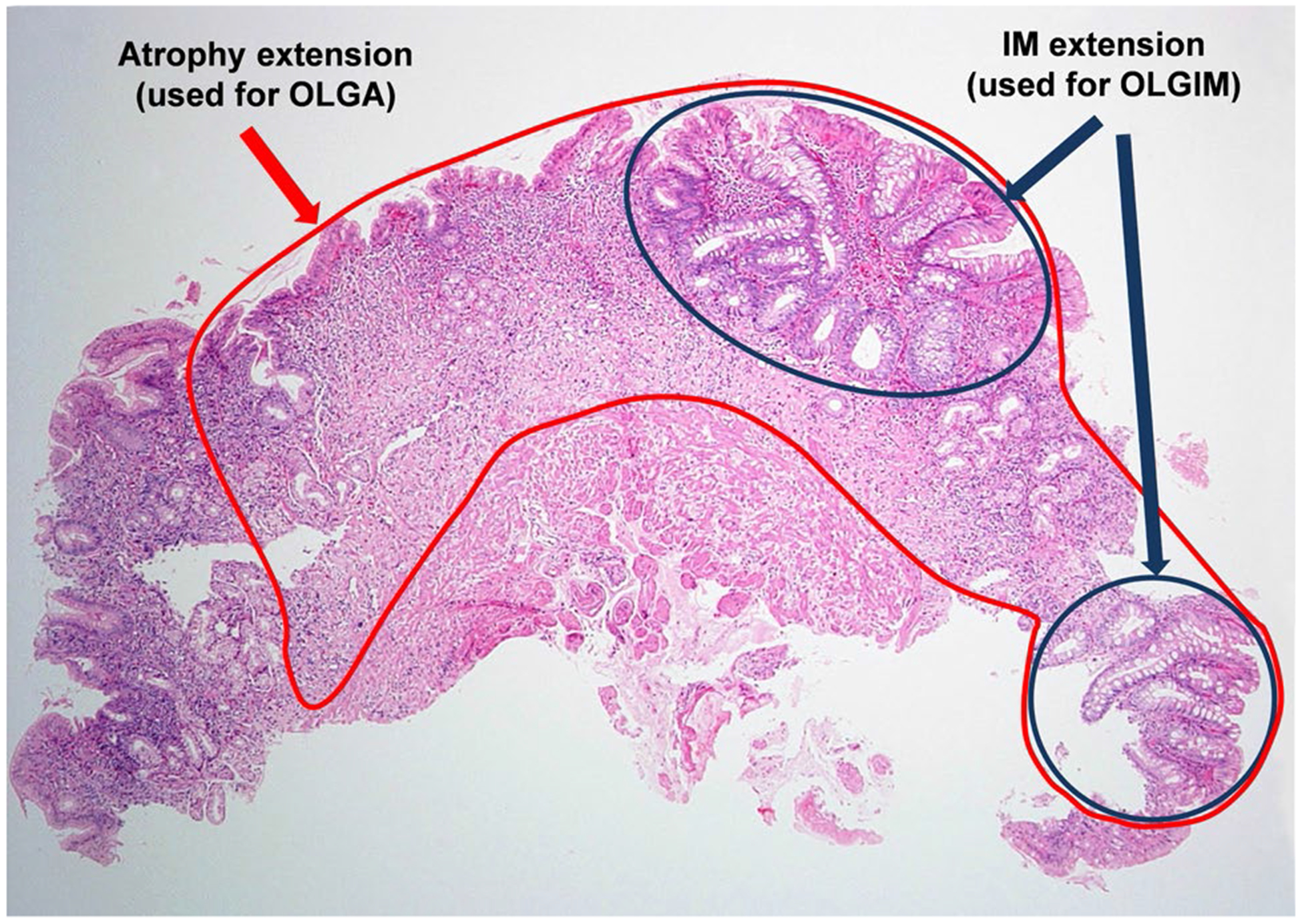
Fig. 1.

Assessment of atrophy at the single biopsy level. In this example, about 90% of the native epithelium is lost (circled in red) and about 30% has been replaced by IM (circled in dark blue). Therefore, there is 90% atrophy (for OLGA) and 30% IM (for OLGIM). Each biopsy is assessed individually. Then, the average of 3 antrum/incisura biopsies and the average of 2 corpus biopsies are used to assign the OLGA and OLGIM stages. For a detailed tutorial for applying OLGA system, see Rugge et al., Dig Liver Dis. 2008;40:650–8
