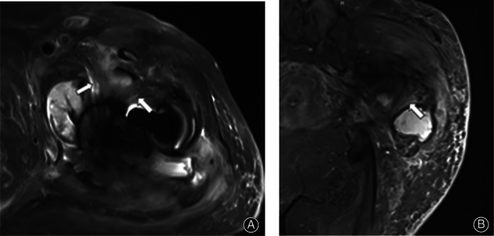
Fig 1.

A 73‐year‐old female patient underwent THA for 4 years. On T2WI‐tirm‐SEMAC, the synovium around the joint in the axial position (A) and coronal position (B) showed LHS (arrow), which could be distinguished from the normal muscle and tendon tissues by anatomical tissue location, travel, and morphology. The patient has a history of diabetes and complained of resting pain in the right hip for >4 months. Admission examination revealed an erythrocyte sedimentation rate of 80 mm/h, C‐reactive protein was 71.81 mg/L, and white blood cells were 8.3 × 10⁹/L. Surgery was performed; a large amount of purulent effusion was found during the operation, and the number of neutrophils was >5/high magnification field of vision. The final diagnosis was PJI.
