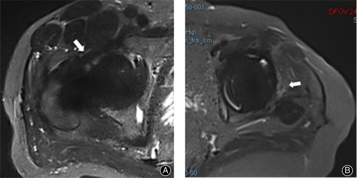
Fig 2.

T2WI‐tirm‐SEMAC axial images of two patients underwent THA after MRI examination. (A) Male, 48‐years‐old, 1 year before THA, the postoperative recovery was better without complaints, and the image was obtained at the time of reexamination. A clear articular cavity boundary was observed in front of the joint, and no LHS image was obtained in the axial position of the joint (arrow). (B) Female, 63‐years‐old, 11 years before THA surgery, hip pain for half a year as the main complaint at the time of admission, diagnosis of aseptic loosening of the prosthesis after THA surgery received revision surgery, arrow indicates light high signal soft tissue shadow, but does not have the lamellated characteristics.
