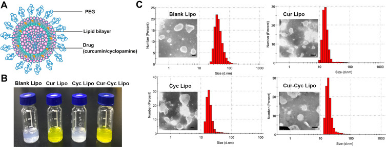
Figure 1.
Characterization of liposomes: (A) schematic of liposomes, (B) photographs of prepared liposomes, and (C) transmission electron micrographs and dynamic light scattering data of the liposomes.
Note: Scale bar, 100 µm. Abbreviations: PEG, polyethylene glycol; Cur, curcumin; Cyc, cyclopamine; Lipo, liposomes.

