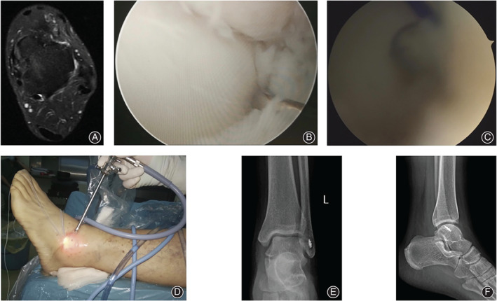
Fig. 5.

A 28‐year‐old male patient with chronic lateral ankle instability for 15 months after failure of 8 months of conservative treatment. (A) The preoperative MRI showed the integrity of the ATFL was interrupted. (B) The intra‐operative view under the arthroscope showed the ATFL detachment at the fibular side, and the laxity of the ATFL was recorded after probe palpation. (C) A suture passer was used to pass the anchor arms through the ATFL and inferior extensor retinaculum. (D) The anchor arms were sutured by horizontal mattress suture fashion. (E) The postoperative anterior–posterior X‐ray film of the operated ankle. (F) The lateral X‐ray film of the left ankle after the surgery.
