
Fig. 1.
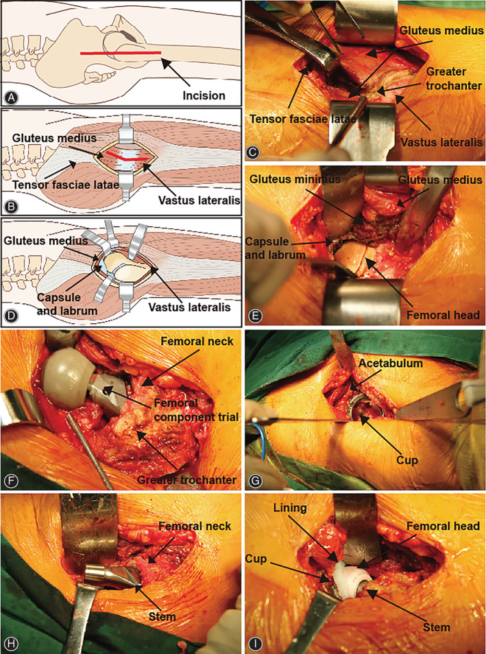
THA using the modified Hardinge approach. (A) The incision was started 3–5 cm proximal to the apex of the greater trochanter and extended distally about 5–7 cm in line with the femur. (B) and (C) The tendon and muscle fibers of the gluteus medius were visualized and split in a one‐third anterior/two‐thirds posterior fashion. (D) and (E) After the gluteus minimus was split, a capsulectomy and labrumectomy was performed to facilitate exposure and dislocation of the hip. (F) The proximal femur was prepared first to determine the anteversion of the stem. (G) The acetabulum was prepared, and the press‐fit cup was fixed on the acetabulum. (H) The proximal femur was further prepared, and the stem was fixed in the proximal femur. (I) The hip was reduced after all of the procedures, and the stability was assessed.