Figure 1.

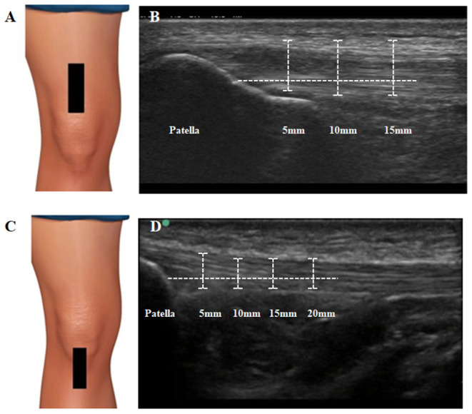
Ultrasound assessment and measurement of quadriceps and patellar tendon. (A) Transducer placement for quadriceps tendon. The transducer was placed in the long axis of this tendon, proximal to the upper edge of the patella. (B) Measurement procedures for quadriceps tendon thickness. Thickness was assessed on three points along the QT located at 5–10–15 mm lateral to the reference point (hyperechoic region of the patella). The QT borders were defined inferiorly as the first hyperechoic region between superficial and deep layers. (C) Transducer placement for patellar tendon. The linear transducer was placed longitudinally distal to the patella. (D) Measurement procedures for patellar tendon thickness. Thickness was assessed on four locations, set at 5–10–15–20 mm inferior to the apex of the patella. Tendon borders were defined inferiorly as the first hyperechoic region between the subcutaneous tissue and the deep fascia layer.
