
FIGURE 3.
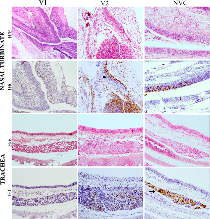
Histopathological lesions and immunohistochemical detection of influenza virus nucleoprotein in nasal turbinates and trachea at 5 dpi. Haematoxylin and eosin (HE) staining of tissues and immunohistochemical (IHC) detection of influenza virus nucleoprotein (NP) showed a moderate fibrinosuppurative rhinitis and lymphoplasmacytic tracheitis observed in group V2, characterised by increased number of inflammatory infiltrates and epithelial damage than the V1 (vaccine 1) group and the non‐vaccinated control (NVC) group. Viral NP immunolabelling observed in respiratory and olfactory epithelium (arrowhead) and glandular epithelium (arrow) of the trachea and nasal turbinates in all groups. Magnification 10x