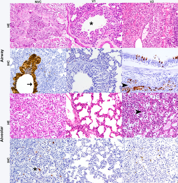
FIGURE 5.

Lung histopathological lesions and viral antigen immunohistochemistry (IHC) in airway and alveolar areas at 3 days post‐infection (dpi). Haematoxylin and eosin (HE) stain revealed that lesions ranged from absent (star) to minimal bronchiolitis in the vaccine 1 (V1) group to more severe lesions observed in the vaccine 2 (V2) group and the non‐vaccinated control (NVC) group. Lesions in the V2 and NVC groups evaluated in HE stain were similar and were consistent with a mild to moderate bronchointerstitial pneumonia characterised by airway and alveolar epithelial necrosis accompanied by inflammatory infiltrates filling the alveolar spaces (arrowhead) and airways. Magnification 20×. Increased numbers of viral nucleoprotein immunolabelled cells were observed by IHC in the V2 and NVC groups in comparison with the V1 group. Positive immunolabelling by IHC was observed in the airway (arrow), glandular epithelium (arrowhead) and the alveolar epithelium (star) of NVC and V2 group ferrets. Magnification 20x
