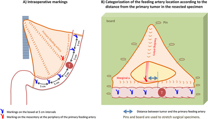
Figure 3.

Intraoperative markings and identification of feeding arteries in resected specimens. Intraoperative markings on the bowel at 5 cm intervals make 11 pericolic segments: i.e. primary tumor area; 0 < D ≤ 5 cm (proximal/distal sides); 5 < D ≤ 10 cm (proximal/distal sides); 10 < D ≤ 15 cm (proximal/distal sides); 15 < D ≤ 20 cm (proximal/distal sides) and 20 cm < D (proximal/distal sides). The location of feeding arteries and pericolic lymph nodes are classified according to these segments.
