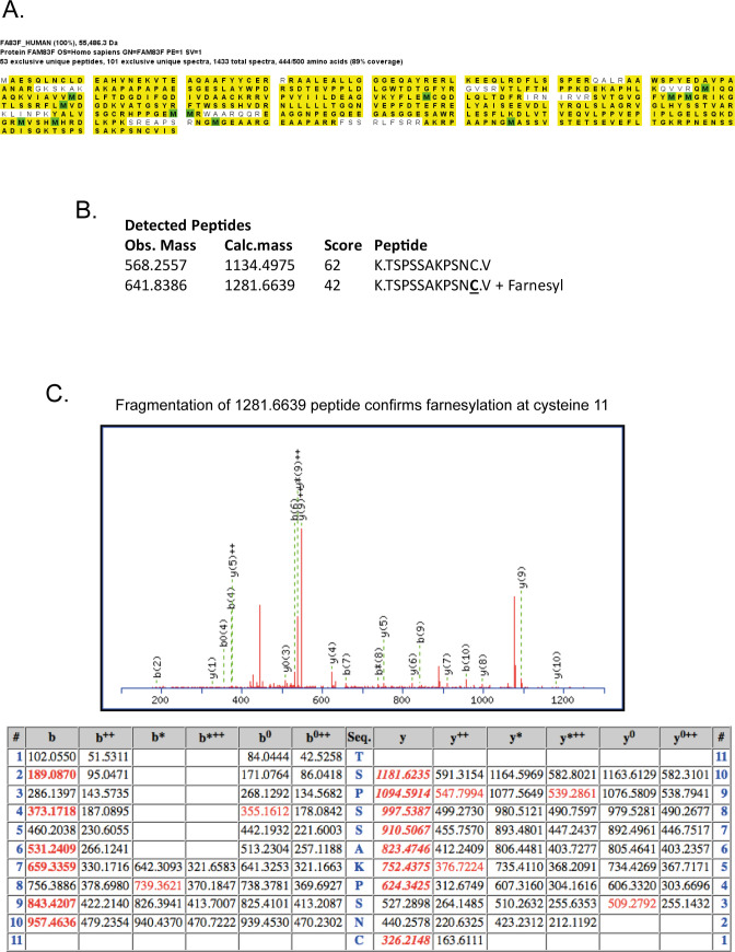
Figure S3. FAM83F is identified as farnesylated by mass spectrometry.
(A) HEK-293 Flp/Trx cells expressing GFP-FAM83F were lysed and subjected to immunoprecipitation with GFP trap beads. GFP eluted proteins were resolved by SDS–PAGE, protein bands were excised and trpysin-digested for mass spectrometry analysis. Mass spectrometry peptide coverage of the FAM83F protein sequence as demonstrated by highlighted residues. (B) Two detected peptides which correspond to the C′-terminal residues of FAM83F protein with both non-farnesylated peptides and farnesylated peptides identified by mass spectrometry. (C) Fragmentation of the farnesylated peptide to confirm presence of a farnesyl group on the cysteine residue which corresponds to the CAAX box cysteine.

